
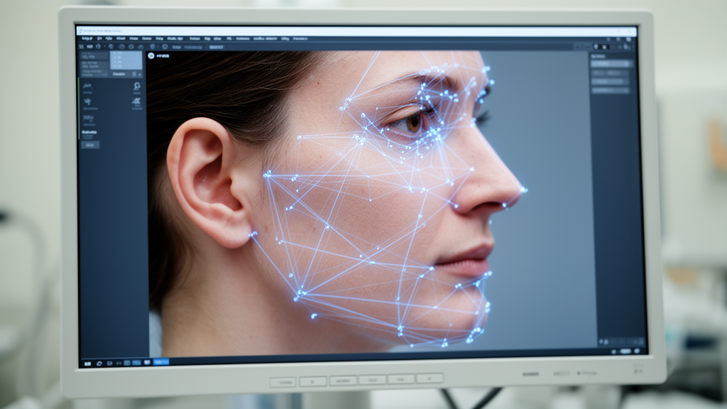
Orthodontic diagnosis and treatment planning have historically relied on lateral cephalometric radiographs—introduced back in 1931—to assess dental and skeletal malocclusions, track adolescent growth, and evaluate treatment progress. However, manual landmark identification on these X-rays is time-intensive, demands high professional skill, and is prone to both inter-observer and intra-observer variability. Furthermore, traditional imaging exposes patients to ionizing radiation.
To overcome these hurdles, a research team has developed a novel deep learning computational framework. This system successfully predicts lateral cephalometric measurements directly from standard lateral facial photographs, offering a reliable, radiation-free alternative for clinical assessment.
Data Collection and Study Methodology
To train and evaluate the artificial intelligence model, researchers gathered pretreatment clinical records from 1,500 patients with diverse malocclusions. The data was collected from patients treated at the Faculty of Dentistry at Cairo University between October 2020 and October 2024.
The extensive dataset was organized into two distinct groups:
- Development Set: Comprising 1,350 patient records utilized for training and validation phases.
- Test Set: Comprising 150 independent patient records that remained completely concealed throughout training and validation.
To establish the ground truth, experts performed manual landmark identification for 19 skeletal, dental, and soft tissue landmarks using Dolphin imaging software. Corresponding lateral profile photographs, captured under standardized lighting and positioning, underwent resizing, rescaling, and an image superimposition protocol that accurately transferred the cephalometric tracings onto the facial images. The dataset was then subjected to normalization and data augmentation preprocessing steps to bolster robustness against variations in patient positioning or photographic technique.
Dual-Task Architecture and Technical Design
The technical framework utilizes a two-stage ensemble system engineered to maximize landmark prediction precision. The primary detector, HRNetV2, simultaneously executes two essential tasks:
- Landmark Coordinate Regression: Focuses on local precision.
- Skeletal Pattern Classification: Based on the ANB angle, providing crucial global morphological understanding to inform local landmark localization.
To further enhance accuracy, developers integrated an adaptive multilayer perceptron (MLP) acting as a tutor network. This secondary component identifies error patterns generated by the HRNetV2 detector and outputs refined landmark coordinates. The training process utilized the Adam optimizer guided by a combined loss function that weighted the regression task twice as heavily as the classification task, alongside extra weighting for underrepresented skeletal class III cases.
Performance Accuracy and Clinical Implications
The ensemble-MLP model achieved a mean radial error of 2.317 ± 1.80 millimeters. Success detection rates reached 53.8% at the 2-millimeter threshold and 84.5% at the 4-millimeter threshold. Performance varied across anatomy:
- Soft Tissue Landmarks: Features like the nasal tip, subnasale, and lips showed the lowest localization errors (mean radial error of 1.101 millimeters) due to high visual contrast and distinct contours.
- Posterior Skeletal Landmarks: Deeper, virtual structures such as the Sella, Gonion, and PNS exhibited higher localization errors because of complex definitions and inter-individual anatomical variability.
Automated cephalometric measurements derived from predicted coordinates closely matched ground truth data, yielding a mean absolute error of 1.46 degrees for the ANB angle. A Bland-Altman analysis demonstrated minimal systematic bias, confirming results hold strong clinical agreement with Dolphin-software benchmarks.
While this approach marks a significant technological advancement, researchers emphasize it is meant to enhance rather than replace traditional radiographs. Eliminating radiation exposure makes the tool particularly valuable for preliminary orthodontic screenings, remote evaluations, and frequent longitudinal tracking. Current limitations—such as single-center data collection and unquantified variables like soft tissue thickness—point the way for future research into broader populations and more integrated diagnostic tools.
