
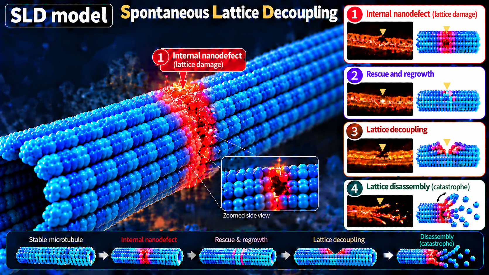
Researchers at Kanazawa University in Japan have uncovered hidden structural vulnerabilities within microtubules—the microscopic tubular networks that support living cells—even after the structures have been treated with a stabilizing cancer therapy. Published in October 2026, the study provides a closer look at how these cellular supports can break down under laboratory conditions.
Observing Nanoscale Changes in Real Time
Led by Richard W. Wong alongside Xin Zhang and colleagues at Kanazawa University's Nano Life Science Institute (WPI-NanoLSI), the research team used high-speed atomic force microscopy (HS-AFM) to examine microtubules assembled from purified tubulin proteins and stabilized with paclitaxel, a medication commonly used to treat breast and ovarian cancers. While conventional imaging methods yield static pictures, HS-AFM allowed the team to scan biological structures in liquid with nanometer-scale detail and observe dynamic changes as they occurred.
Although paclitaxel is designed to stabilize microtubules and interfere with normal cell division, close examination revealed unexpected developments. Small defects, some starting as shallow depressions just a few nanometers across, formed within otherwise intact regions of the microtubule wall. These wall defects frequently determined whether a microtubule remained intact or collapsed, leading the team to propose a new model called Spontaneous Lattice Decoupling (SLD).
Four Distinct Patterns of Breakdown
By analyzing 73 individual collapse events, the researchers identified four distinct ways microtubules break down:
- 42 percent began at one end of the microtubule, aligning with conventional models.
- 40 percent began at internal wall defects, spreading in both directions from the damaged area.
- 10 percent involved simultaneous breakdown from both ends.
- 8 percent originated at multiple internal sites simultaneously.
The findings indicate that internal structural defects serve as a substantial source of microtubule instability, suggesting that stability depends heavily on the condition of the protein framework along the entire length of the structure rather than exclusively at its ends.
Study Context and Limitations
The authors noted important limitations to their work. The experiments were conducted on purified proteins under laboratory conditions rather than inside living cells, and the paclitaxel concentrations used were chosen to provide strong stabilization during prolonged imaging rather than to mimic exact clinical drug exposure in patients. Further research is necessary to determine whether these internal structural processes occur inside living cells and if they directly influence cancer drug sensitivity or resistance.
